Module 2: Plants and Essential Oils PDF
Module 2: Plants and Essential Oils PDF – A4 paper
Foundations of Essential Oil Therapy
MODULE ONE PDF
Dilution Charts
Essential Oil Dilution Chart
| Essential Oil Dilution Chart | ||||||
|---|---|---|---|---|---|---|
| Carrier oil in ounces | .5% dilution | 1% | 2.5% | 3% | 5% | 10% |
| ½ ounce | 1-3 drops | 3-5 drops | 8-11 drops | 9-13 drops | 15-23 drops | 30-46 drops |
| 1 ounce | 3-5 drops | 6-9 drops | 15-23 drops | 18-27 drops | 30-45 drops | 60-90 drops |
| 2 ounces | 6-10 drops | 12-18 drops | 30-46 drops | 36-54 drops | 60-90 drops | 120-180 drops |
| 4 ounces | 12-20 drops | 24-36 drops | 60-92 drops | 72-108 drops | 120-180 drops | 240-360 drops |
| **This chart includes the traditional dosage plus changes in drops by Tisserand and Young (2013) which tended to be higher than the traditional dosage as taught by most of us in the world of aromatherapy. | ||||||
The dilution you use should be based on the same factors you consider in your selection of essential oils: the client’s constitution and age, the condition being addressed (acute or chronic), energetics, assessments (e.g., tongue or pulse), the time frame for treatment, and the client’s goals.
Essential Oil Dilution Rates and Indications
| Dilution Rate and Indications | |
|---|---|
| Dilution in % | Purpose and Indications |
| 0 | Essential oils should not be used for infants under 6 months of age unless absolutely necessary. |
| 0.25% - 0.5% | Infants 6 months or older, frail or elderly individuals, immune compromised individuals |
| < 3% | Sensitive and fragile mucus membranes, e.g. nasal or auricular applications. Repairing action on the external layers of the skin |
| 1% | Children 2-5 years old, pregnancy; facial creams |
| 1.5% | Subtle aromatherapy, emotional and energetic work, pregnancy, frail or elderly individuals, facial creams and lotions, exfoliants |
| Between 2 - 5% | Action on the nervous system, emotional well-being, and response to daily stress. Action on the endocrine system. Holistic aromatherapy, general massage work, general skincare, massage oils, lotions, facial oils, body oils, and body butters. |
| 7% | Action on venous and lymphatic circulation. General strength for most blends. This is a stronger dilution that can be used for treatment massage and localized treatment work, wound healing, body oils and butters, and salves. |
| 10% | Action on the joints, tendons, muscles and inflammation. This is a stronger dilution for muscular aches and pains, trauma injury, treatment massage, acute physical pain, localized treatment work, and salves. skin infection/dermatophytes |
| 20% | Similar indications to 10% or 30%. This is the highest dilution rate recommended for dermacaustic essential oils (essential oils that are rich in phenols and aldehydes) |
| 30% | Powerful local action. Warts, acute trauma to the musculoskeletal system, skin infection/dermatophytes |
| 30 - 50% | For acute treatments with non-aggressive essential oils |
| Undiluted | For localized frictions. Antispasmodic (e.g. muscles, coughs), expectorant, antimicrobial, warming and acute trauma with non-aggressive essential oils |
Lesson 5: The Cardiovascular System A&P
Introduction
The cardiovascular system, also known as the circulatory system, is a remarkable network of organs and blood vessels that delivers vital nutrients and oxygen to all the cells in the body while removing metabolic waste products. It’s like a complex highway system with the heart as the central pump that keeps traffic flowing. Every single cell in the body depends on the cardiovascular system to survive and function optimally. Let’s embark on a journey to explore the anatomy and physiology of this fascinating system.
The Heart: The Powerhouse of the Cardiovascular System
At the center of the cardiovascular system lies the heart, a fist-sized muscular organ that tirelessly pumps blood throughout the body. The heart is situated in the chest cavity, nestled between the lungs and protected by the rib cage. This incredible pump is divided into four chambers – two atria and two ventricles. The right side of the heart receives deoxygenated blood from the body and pumps it to the lungs, while the left side collects oxygenated blood from the lungs and propels it to the rest of the body.
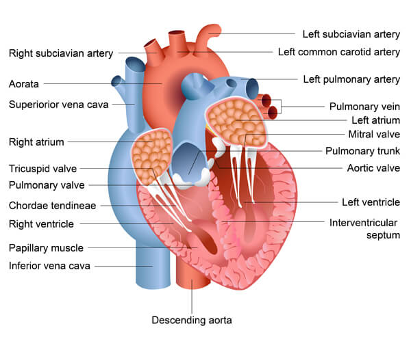
The walls of the heart are composed of three layers: the epicardium (outer layer), myocardium (muscular middle layer) and endocardium (inner layer). The myocardium is responsible for the heart’s pumping action. It is made up of specialized cardiac muscle cells called myocytes that contract and relax in a coordinated manner. These contractions are triggered by electrical impulses that originate in the heart’s own conduction system.
The heart’s conduction system consists of specialized cells that spontaneously generate electrical signals. The sinoatrial (SA) node, located in the right atrium, is the natural pacemaker of the heart. It initiates each heartbeat by sending an electrical impulse that spreads through the atria, causing them to contract. The signal then reaches the atrioventricular (AV) node, which delays it slightly, allowing the ventricles to fill with blood before contracting. Finally, the impulse travels down the bundle of His and Purkinje fibers, triggering a coordinated contraction of the ventricles that forcefully pumps blood out of the heart.
Blood Vessels: The Body’s Highway System
Blood vessels form an extensive network that carries blood to and from the heart and throughout the body. There are three main types of blood vessels: arteries, capillaries, and veins. Arteries carry oxygen-rich blood away from the heart to the body’s tissues. They have thick, muscular walls to withstand the high pressure of blood pumped out of the heart. As arteries branch into smaller vessels called arterioles, they begin to deliver blood to the capillaries.
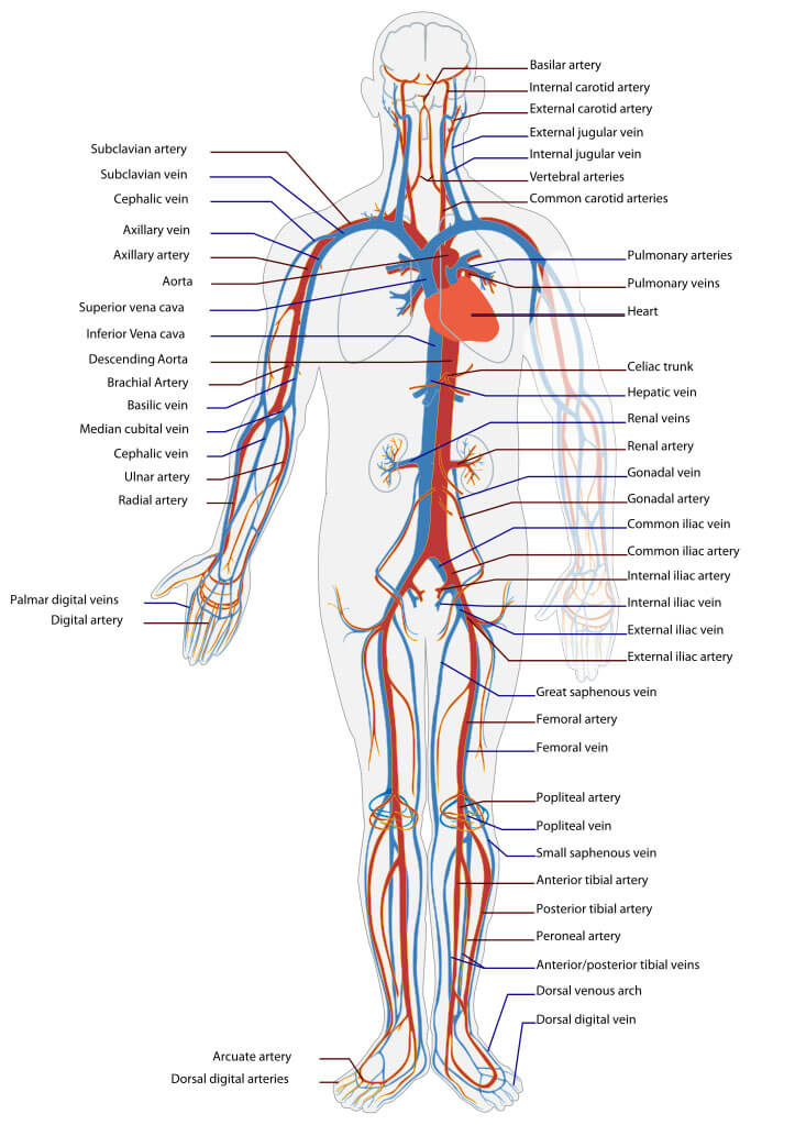
Capillaries are the tiniest and most numerous of the blood vessels. These thin-walled, microscopic tubes allow for the exchange of nutrients, oxygen, and waste products between the blood and individual cells. Capillaries then merge to form venules, which are small veins that carry deoxygenated blood back towards the heart. As venules unite, they become larger veins with thinner walls and valves to prevent blood from flowing backward. Veins rely on surrounding muscle contractions to help push blood back to the heart.
The blood vessels are lined with a thin layer of cells called the endothelium. The endothelium plays a crucial role in regulating blood flow, blood clotting, and the exchange of materials between the blood and tissues. It also secretes important substances like nitric oxide, which helps blood vessels dilate and maintains their health.
The Conducting Circuits: Systemic and Pulmonary Circulation
The cardiovascular system consists of two main circuits: the systemic circulation and the pulmonary circulation. The systemic circulation carries oxygenated blood from the left side of the heart to all the tissues of the body (except the lungs) and returns deoxygenated blood to the right side of the heart. The pulmonary circulation transports deoxygenated blood from the right side of the heart to the lungs, where it picks up oxygen and releases carbon dioxide. The freshly oxygenated blood then returns to the left side of the heart to be pumped out into the systemic circulation.
The heart also has its own blood supply through the coronary circulation. The coronary arteries branch off the aorta (the main artery leaving the heart) and supply the heart muscle with oxygen-rich blood. If these arteries become blocked or narrowed, it can lead to a heart attack.
The Lifeblood: Composition and Functions of Blood
Blood is a specialized connective tissue that consists of cells suspended in a liquid matrix called plasma.
It has four main components: red blood cells (erythrocytes), white blood cells (leukocytes), platelets (thrombocytes), and plasma.
Red blood cells are the most numerous blood cells and give blood its characteristic red color. They are packed with hemoglobin, an iron-rich protein that binds to oxygen in the lungs and delivers it to the body’s tissues. White blood cells are part of the immune system and help defend the body against infections and diseases. Platelets are small cell fragments that play a crucial role in blood clotting, which stops bleeding when blood vessels are damaged.
Plasma is a straw-colored liquid that makes up about 55% of blood volume. It consists of water, proteins, electrolytes, nutrients, hormones, and waste products. Plasma helps maintain blood pressure and volume, transports substances throughout the body, and plays a role in heat regulation and pH balance.
Blood has three main functions: transportation, regulation, and protection. It transports oxygen, nutrients, hormones, and waste products throughout the body. It helps regulate body temperature, pH balance, and fluid balance. Blood also protects the body through clotting mechanisms that prevent blood loss and white blood cells that fight infections.
Cardiovascular Health and Homeostatic Imbalances
A healthy cardiovascular system is essential for overall health and well-being. However, various factors can disrupt the delicate balance and lead to cardiovascular diseases (CVDs), which are the leading cause of death worldwide.
Atherosclerosis is a common condition in which plaque builds up inside the arteries, causing them to narrow and harden. This restricts blood flow and increases the risk of heart attack and stroke. High blood pressure (hypertension), high blood cholesterol, diabetes, smoking, obesity, physical inactivity, and unhealthy diet are major risk factors for atherosclerosis and CVDs.
Heart valve disorders can also affect cardiovascular function. The heart valves ensure that blood flows in the correct direction through the heart. If they become damaged or defective, blood may leak backward or have difficulty moving forward, straining the heart and reducing its efficiency.
Arrhythmias are problems with the rate or rhythm of the heartbeat. They occur when the electrical impulses that coordinate the heart’s contractions fire irregularly, causing the heart to beat too fast, too slow, or erratically. Some arrhythmias are harmless, while others can be life-threatening.
Congenital heart defects are structural problems with the heart that are present at birth. They can affect the heart walls, valves, or blood vessels, changing the normal flow of blood through the heart. While some defects are minor and cause no problems, others require medical intervention or surgery.
Maintaining the Health of the Cardiovascular System
“The vitality and tone of the whole circulatory system is fundamental to life and to the integration of all the parts of the body.” (David Hoffman)
By attending to the healthy circulation of blood and lymph in the body, we are encouraging the healthy cleansing of toxins from the body and the healthy exchange of nutrients and oxygen into every cell.
The health of the cardiovascular system depends on:
- Exercise – Regular exercise can support healthy blood and lymph flow throughout the entire body.
- Stress level – Stress impacts circulation, and long-term stress can result in atherosclerosis and other circulatory disorders. Practicing good stress-reducing exercises can drastically reduce the likelihood of developing poor circulation problems.
- Diet – Healthy blood and lymph circulation depends on a good diet low in fat.
- Tobacco and alcohol – Tobacco and alcohol both adversely affect the health of circulation.
Any problems arising with the circulation of blood and lymph in the body should always include a good look at the above four elements within the individual’s life to ensure adequate life changes or wellness additions are also made with aromatherapy treatment. This way, the system is addressed in a holistic way.
Lesson 4: The Female Reproductive System A&P
Introduction
The female reproductive system is a remarkable network of organs that work together to create and sustain new life. From the monthly cycle of ovulation and menstruation to the miraculous process of pregnancy and childbirth, this system is essential for the continuation of the human species. In this lesson, we’ll explore the anatomy and physiology of the female reproductive system, the intricate hormonal dance of the menstrual cycle, and some common conditions that can affect reproductive health. We’ll also discuss the menopausal transition and its impact on a woman’s body and well-being.
The Ovaries: The Powerhouses of Female Reproduction
At the heart of the female reproductive system lie the ovaries, two small, almond-shaped glands that house a woman’s lifetime supply of eggs. These powerhouses not only produce and release eggs for potential fertilization but also secrete the essential female hormones estrogen and progesterone.
Each month, under the influence of follicle-stimulating hormone (FSH) from the pituitary gland, several follicles (fluid-filled sacs containing immature eggs) begin to develop within the ovaries. Usually, only one follicle will mature fully, rupturing to release its egg during ovulation. The remaining follicles degenerate. The released egg is swept into the fallopian tube, where fertilization may occur if sperm are present.
After ovulation, the ruptured follicle transforms into the corpus luteum, a temporary endocrine structure that secretes progesterone to prepare the uterus for potential pregnancy. If fertilization does not occur, the corpus luteum degenerates, causing hormone levels to drop and triggering menstruation.
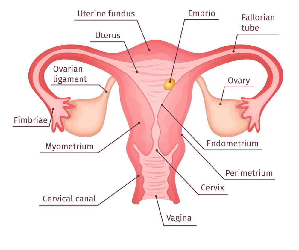
The Uterus and Fallopian Tubes: Nurturing and Transporting New Life
The uterus, a pear-shaped, muscular organ, is the site where a fertilized egg implants and develops into a fetus. The inner lining of the uterus, called the endometrium, thickens each month in preparation for a potential pregnancy. If fertilization does not occur, the endometrium is shed during menstruation.
The fallopian tubes, also called uterine tubes, serve as the pathway for the egg to travel from the ovary to the uterus. Finger-like projections at the end of each tube, called fimbriae, help guide the released egg into the tube. Fertilization typically occurs in the fallopian tubes, and the fertilized egg, now called a zygote, continues its journey to the uterus for implantation.
The Menstrual Cycle: A Delicate Hormonal Balance
The menstrual cycle is a complex interplay of hormones that prepares the female body for potential pregnancy each month. The cycle is regulated by the hypothalamus and pituitary gland in the brain, which send signals to the ovaries to produce estrogen and progesterone.
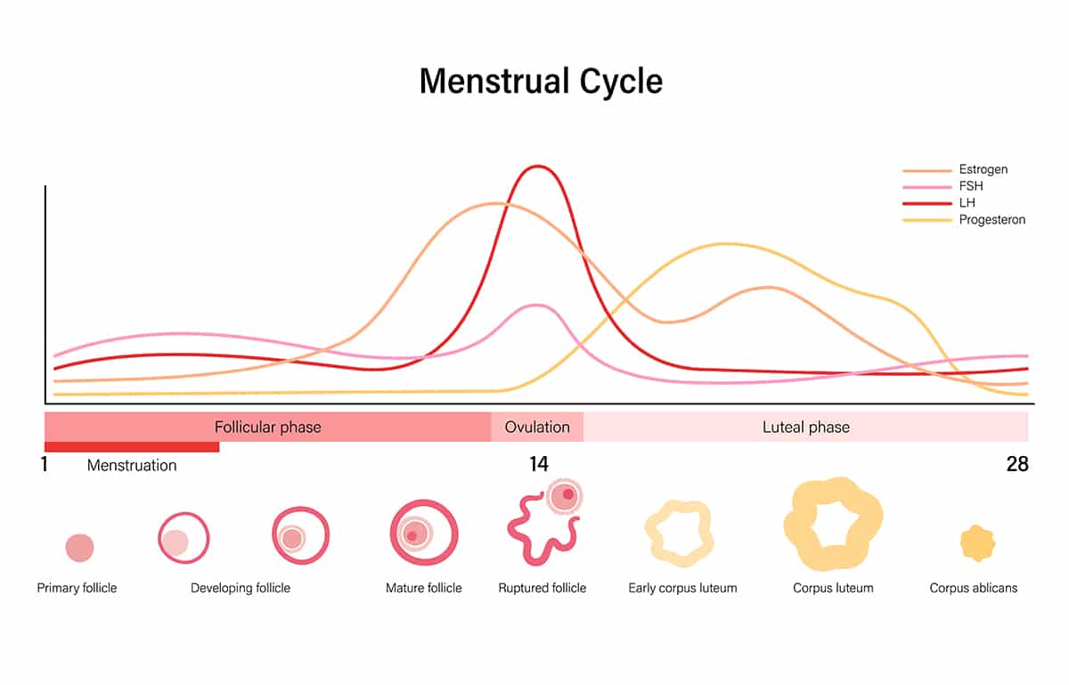
A typical menstrual cycle lasts about 28 days and can be divided into four phases:
- Menstrual phase: The endometrium is shed if pregnancy has not occurred, resulting in menstrual flow.
- Follicular phase: Estrogen levels rise, causing the endometrium to thicken and a new egg to mature in the ovary.
- Ovulation: A surge of luteinizing hormone (LH) triggers the release of the mature egg from the ovary.
- Luteal phase: The corpus luteum produces progesterone to maintain the endometrium. If pregnancy does not occur, the corpus luteum disintegrates, hormone levels drop, and the cycle begins again.
While this cyclical process is a normal part of female physiology, it can sometimes cause physical and emotional symptoms, such as cramps, bloating, and mood changes, known as premenstrual syndrome (PMS).
Vagina, Cervix, and External Genitalia: Gateways to the Reproductive System
The vagina is a muscular canal that connects the uterus to the outside world. It serves as a passageway for menstrual flow, receives the penis during sexual intercourse, and acts as the birth canal during childbirth. The vaginal walls are lined with a mucous membrane that helps maintain a healthy balance of bacteria.
The cervix is the lower, narrow portion of the uterus that extends into the vagina. It produces mucus that can either facilitate or block the passage of sperm, depending on the stage of the menstrual cycle. The cervix also dilates during childbirth to allow the baby to pass through.
The external genitalia, collectively called the vulva, include the labia (outer and inner lips), clitoris, and openings of the urethra and vagina. These structures play important roles in sexual arousal, pleasure, and protection of the internal reproductive organs.
Menopause: A Time of Transition
Menopause marks the end of a woman’s reproductive years, typically occurring in the late 40s or early 50s. During this time, the ovaries gradually produce less estrogen and progesterone, causing menstrual cycles to become irregular and eventually stop altogether.
The menopausal transition can bring a range of symptoms, such as hot flashes, night sweats, vaginal dryness, mood changes, and sleep disturbances. These symptoms are caused by the body’s adjustment to lower hormone levels. While some women may experience only mild symptoms, others may have significant discomfort that impacts their quality of life.
After menopause, women are at increased risk for certain health conditions, such as osteoporosis and heart disease, due to the protective effects of estrogen. However, lifestyle changes and medical treatments can help manage these risks.
Common Reproductive Health Concerns
Like any complex system, the female reproductive system is susceptible to various disorders and diseases. Some common issues include:
- Polycystic ovary syndrome (PCOS): A hormonal disorder that causes enlarged ovaries with small cysts, irregular periods, and high levels of male hormones, which can lead to acne, excess hair growth, and fertility issues.
- Endometriosis: A condition in which tissue similar to the uterine lining grows outside the uterus, causing painful periods, pelvic pain, and sometimes infertility.
- Uterine fibroids: Non-cancerous growths in the uterus that can cause heavy menstrual bleeding, pelvic pain, and pressure on nearby organs.
- Cervical and ovarian cancer: Malignancies of the cervix and ovaries that can be detected through regular screenings, such as Pap smears and pelvic exams.
- Infertility: Difficulty conceiving due to various factors, such as hormonal imbalances, blocked fallopian tubes, or age-related decline in egg quality and quantity.
Summary
The female reproductive system is a testament to the incredible complexity and resilience of the human body. From the intricate dance of hormones that orchestrate the menstrual cycle to the remarkable journey of egg and sperm uniting to create new life, this system is essential for the continuation of our species. While it can be a source of great joy and fulfillment, it can also present unique challenges and health concerns throughout a woman’s life.
By understanding how the female reproductive system works and taking steps to maintain its health, women can empower themselves to navigate the many stages of their reproductive journey with confidence and grace. As we marvel at the intricacies of the female body, let us also remember to approach reproductive health with compassion, respect, and an appreciation for each woman’s diverse experiences and choices.
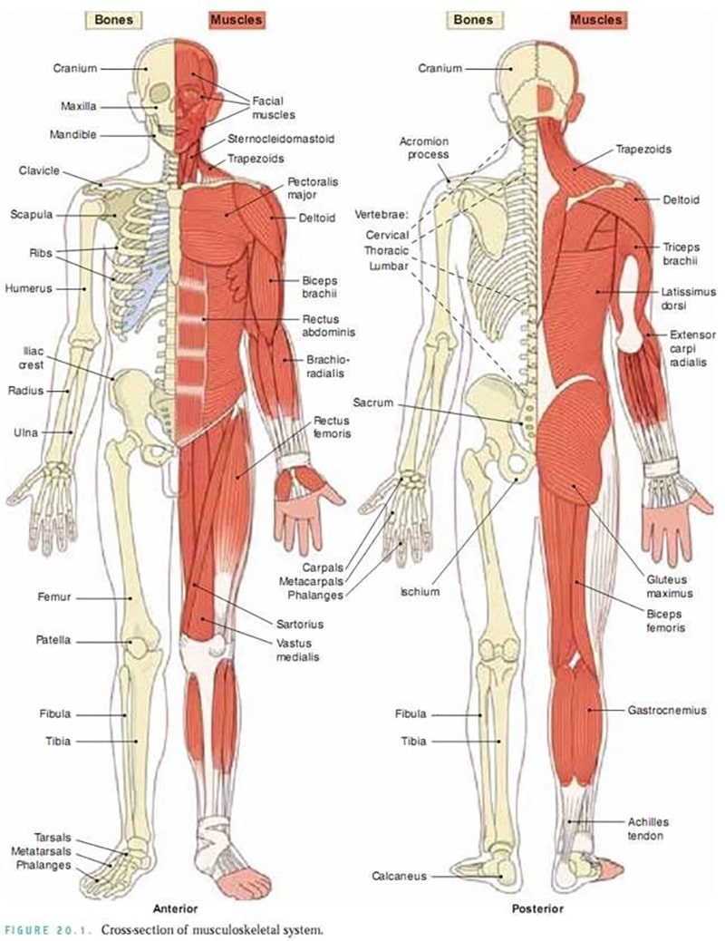
Lesson 3: The Musculoskeletal System A&P
Introduction
The musculoskeletal system is a fascinating component of the human body that enables us to move, provides support and protection, and performs other crucial functions. This system includes the bones, muscles, tendons, ligaments, and other connective tissues working together in a beautifully orchestrated way. Let’s dive in and explore the key parts and functions of the musculoskeletal system.
The Skeleton: A Strong and Adaptive Framework
At the core of the musculoskeletal system is the skeleton, composed of over 200 bones. The skeleton serves as a framework that supports the body, protects vital organs, provides attachment points for muscles, and even produces blood cells within the bone marrow. Bones come in different shapes and sizes, from the long bones in the arms and legs to the flat bones that make up the skull.
Bones are made of strong and rigid material, yet also exhibit flexibility to withstand impacts. The outer layer of bone, called compact bone, is very dense and sturdy. Inside many bones is spongy bone, a honeycomb-like network that helps reduce weight while maintaining strength. Bones contain cells called osteoblasts that build new bone, osteoclasts that break down old bone, and osteocytes that maintain bone structure. This allows the skeleton to continually remodel and repair itself.
Mighty Muscles: Strength and Movement
Attached to the bones are over 600 muscles that generate movement. Muscles are made up of stretchy fibers that contract and relax. The muscle fibers are bundled together, with each muscle anchored to bones via tendons. When a muscle contracts, it pulls on the bones, causing movement at a joint. Different muscles work in pairs – as one muscle contracts, the opposing muscle relaxes. This coordinated push-pull mechanism allows for smooth, controlled movements.
There are three main muscle types:
- Skeletal muscles are consciously controlled and provide movement, maintain posture, and generate body heat. These are the muscles you use to walk, lift objects, smile, and more.
- Smooth muscles are found in the walls of organs and blood vessels. They work automatically without conscious control to push substances like food through the digestive tract and regulate blood flow.
- Cardiac muscle makes up the heart and pumps blood throughout the body with an automatic rhythm.
All muscle movement requires energy in the form of ATP. When oxygen is available, muscles can generate ATP through aerobic respiration. For quick, intense movements, muscles can also produce ATP anaerobically, although this leads to lactic acid buildup and muscle fatigue. With proper rest, muscles will rebuild their energy stores and repair any tissue damage.
Connective Tissue: The Binding Elements
Connective tissue holds everything together and allows the musculoskeletal system to function properly. This includes ligaments that connect bones to other bones and stabilize joints. For example, the anterior cruciate ligament (ACL) helps secure the knee joint. Tendons are another type of connective tissue that anchors muscles to bones, like the Achilles tendon at the back of the heel.
Cartilage is a firm, rubbery connective tissue that cushions bones at joints and provides a smooth surface for bones to glide over, reducing friction. It also gives shape and support to various body structures, like the nose and ears. Because cartilage has a limited blood supply, injuries are slow to heal.
Fascia is a thin casing of connective tissue that surrounds and stabilizes muscles and organs. Recent research suggests the fascia helps muscles communicate with each other and plays an essential role in coordinating movement. Keeping fascia healthy through stretching and exercise supports overall musculoskeletal function.
Joints: Artfully Articulated
Joints are where two or more bones come together, and they allow for different types and degrees of movement. Some joints, like those between the bones of the skull, allow for very little movement. Others, like the ball-and-socket joint of the shoulder, enable a wide range of motion.
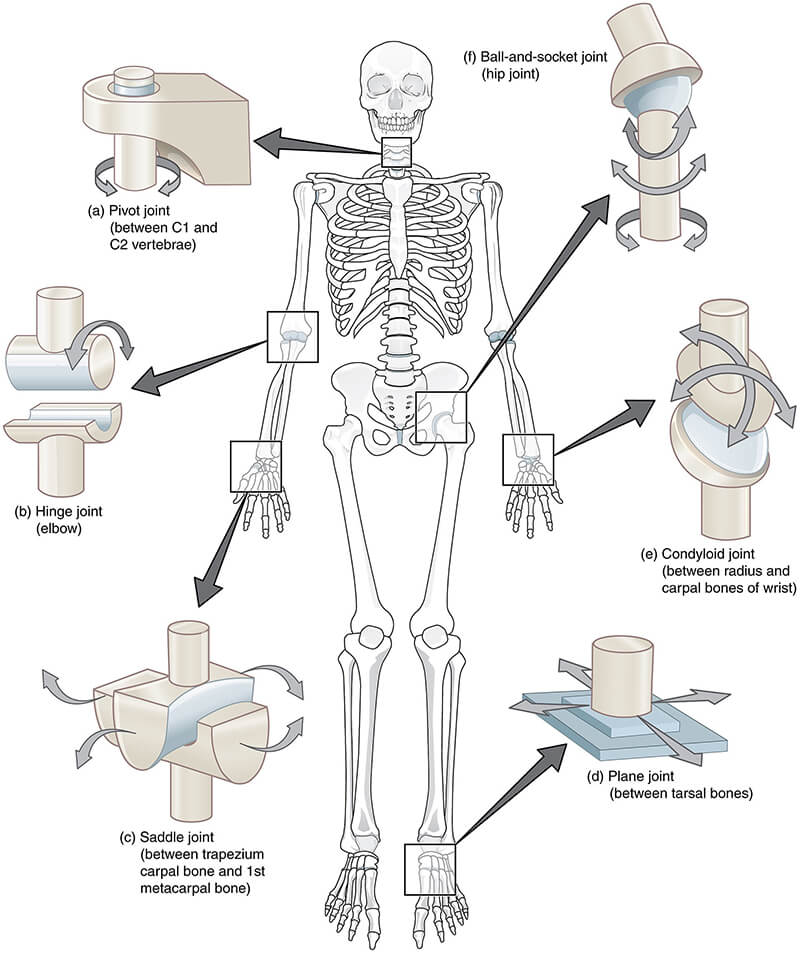
The most common and movable type of joint is the synovial joint. These joints contain a capsule surrounding the bones filled with lubricating synovial fluid. This allows for free movement while keeping the bones from grinding against each other. Cartilage covers the ends of the bones for additional padding and smooth gliding. Ligaments surround the joint to provide stability and prevent excessive movement.
Caring for Your Musculoskeletal Health
Proper nutrition and regular exercise are key to keeping the musculoskeletal system functioning at its best. Bones and muscles require adequate levels of minerals like calcium and magnesium, plus vitamin D to absorb calcium. Weight-bearing exercises stimulate bones to take in more minerals and become stronger. Resistance training stresses muscles, signaling them to build new fibers, resulting in increased strength and tone.
Rest is equally important, as it allows bones, muscles, and connective tissues time to recover and repair any micro-tears. Overuse can lead to injuries like stress fractures, torn ligaments, and strained muscles. Listening to the body’s signals and avoiding excessive strain helps prevent injury.
Musculoskeletal Conditions and Injuries
Like any system of the body, things can sometimes go wrong with bones, muscles, and connective tissues. With aging, bones naturally lose some mass and strength, which can progress to osteoporosis and increased fracture risk if left unchecked. Arthritis is another common condition characterized by joint inflammation and deterioration.
Acute injuries like ligament tears, bone fractures, and muscle strains also frequently occur, often due to falls, collisions, and athletic activities that stress tissues beyond their limits. While painful, most of these injuries heal well if rested and rehabilitated properly. More severe trauma may require surgical repair.
Lesson 2: The Digestive System A&P
Introduction
The digestive system is a remarkable network of organs that transforms the food we eat into the nutrients our bodies need to function, grow, and repair themselves. It’s like a complex processing plant that breaks down raw materials (food) into usable components (nutrients) while discarding the waste. Every cell in our body depends on the digestive system to provide the fuel it needs to carry out its functions.
In this lesson, we’ll take a journey through the digestive tract, exploring the anatomy and physiology of each organ and the role it plays in the process of digestion. We’ll also discuss some common digestive disorders that can disrupt this vital system.
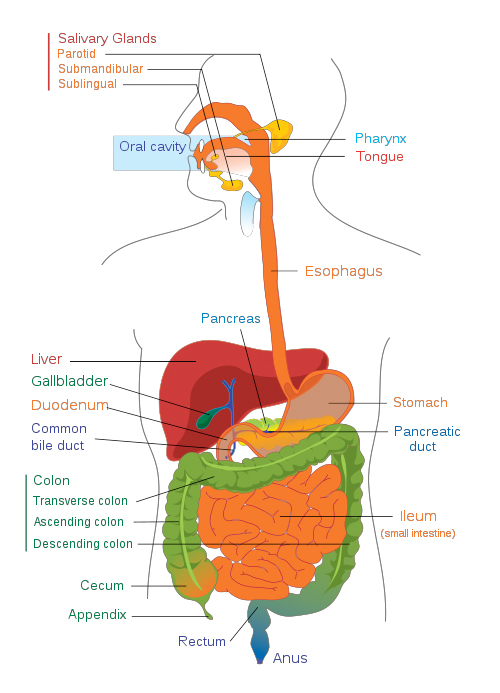
The Journey Begins: The Mouth and Salivary Glands
Digestion begins before we even take our first bite. The sight, smell, or even thought of food triggers the cephalic phase of digestion, stimulating the secretion of saliva and digestive juices. As we chew, our teeth mechanically break down food into smaller pieces while our tongue mixes it with saliva. Saliva contains enzymes like salivary amylase that begin the chemical breakdown of carbohydrates. It also lubricates the food, making it easier to swallow.
The salivary glands, including the parotid, submandibular, and sublingual glands, secrete saliva in response to stimulation from the autonomic nervous system. The parasympathetic nervous system, activated when we are relaxed, stimulates saliva production. Conversely, the sympathetic nervous system, activated during stress, slows or stops saliva secretion. This is why our mouth feels dry when we are anxious or scared.
Down the Hatch: The Pharynx and Esophagus
Once the food is chewed and mixed with saliva, it is swallowed and passes through the pharynx and esophagus on its way to the stomach. The pharynx, a muscular tube connecting the mouth and esophagus, also plays a role in respiration. During swallowing, specialized muscles and cartilage work together to prevent food from entering the trachea and lungs.
The esophagus, a long muscular tube, connects the pharynx to the stomach. It uses rhythmic muscle contractions called peristalsis to push food toward the stomach. At the junction of the esophagus and stomach lies the lower esophageal sphincter (LES), a ring of muscle that opens to allow food into the stomach and then closes to prevent stomach acid from backing up into the esophagus.
The Stomach: A Mixing and Digesting Powerhouse
The stomach is a J-shaped, muscular organ that acts as a temporary storage tank for food. It has three main functions:
- Storage: The stomach can expand to hold up to a liter or more of food and liquid.
- Mixing: The stomach’s strong muscular walls churn and mix food with digestive juices, turning it into a semi-liquid mixture called chyme.
- Digestion: The stomach secretes hydrochloric acid and digestive enzymes like pepsin that break down proteins.
The stomach lining is protected from its own acid by a thick layer of mucus secreted by specialized cells. The stomach also releases hormones like gastrin that stimulate digestive secretions and motility.
Chyme is gradually released from the stomach into the small intestine through the pyloric sphincter.
The Small Intestine: Nutrient Absorption Central
The small intestine is where most digestion and absorption takes place. Despite its name, it is actually the longest part of the digestive tract, measuring about 20 feet in length. It is divided into three sections: the duodenum, jejunum, and ileum.
In the duodenum, chyme from the stomach mixes with digestive secretions from the pancreas, liver, and gallbladder. The pancreas secretes enzymes that break down all major nutrient types – carbohydrates, proteins, and fats. The liver produces bile, which is stored and concentrated in the gallbladder. Bile helps emulsify fats, making them easier for enzymes to break down.
The inner surface of the small intestine is covered with finger-like projections called villi and even smaller projections called microvilli. This highly folded surface greatly increases the surface area for nutrient absorption. Each villus has its own blood and lymphatic vessels that carry absorbed nutrients to the rest of the body.
The Colon: Recycling and Waste Removal
The colon, also known as the large intestine, is the final stop in the digestive tract. Its main functions are to absorb water and electrolytes from the remaining indigestible food matter and to form and eliminate feces.
The colon is home to trillions of bacteria that make up the gut microbiome. These beneficial bacteria ferment undigested fibers, produce certain vitamins (like vitamin K), and support our immune system. Maintaining a healthy balance of gut bacteria is crucial for overall health.
Feces are stored in the rectum until they are eliminated from the body through the anus during defecation. The internal and external anal sphincters control this process.
Common Digestive Disorders
While the digestive system is designed to function efficiently, various factors can disrupt its balance, leading to digestive disorders. Some common issues include:
- Gastroesophageal reflux disease (GERD): When the LES doesn’t close properly, stomach acid can back up into the esophagus, causing heartburn and damaging the esophageal lining.
- Peptic ulcers: Sores that develop in the lining of the stomach or duodenum, often caused by a bacterial infection (H. pylori) or long-term use of certain medications.
- Inflammatory bowel diseases (IBD): Chronic inflammation of the digestive tract, including Crohn’s disease and ulcerative colitis. Symptoms include abdominal pain, diarrhea, and weight loss.
- Irritable bowel syndrome (IBS): A functional disorder characterized by abdominal pain, bloating, and changes in bowel habits (diarrhea or constipation).
- Celiac disease: An autoimmune disorder triggered by gluten, a protein found in wheat, barley, and rye. It damages the villi in the small intestine, impairing nutrient absorption.
Maintaining Digestive Health
To keep your digestive system functioning optimally, focus on these key aspects of digestive health:
- Eat a balanced diet rich in fruits, vegetables, whole grains, and lean proteins.
- Stay hydrated by drinking plenty of water throughout the day.
- Manage stress through relaxation techniques, as stress can disrupt digestion.
- Exercise regularly to maintain a healthy weight and promote regular bowel movements.
- Limit processed foods, excessive alcohol, and tobacco use, which can irritate the digestive tract.
- Listen to your body’s hunger and fullness cues, and eat mindfully.
By understanding how the digestive system works and taking steps to support its health, you can ensure that your body gets the nutrients it needs to thrive.
Summary
The digestive system is a complex and fascinating network of organs working together to break down food, absorb nutrients, and eliminate waste. From the moment we take our first bite to the final stage of elimination, the digestive tract performs a series of mechanical and chemical processes that are essential for our survival. While we may not give much thought to our digestive system when it’s functioning well, digestive disorders can quickly remind us of its importance.
By making lifestyle choices that support digestive health, we can help keep this vital system running smoothly, ensuring that our bodies have the fuel they need to thrive. So, the next time you sit down for a meal, take a moment to appreciate the amazing journey your food is about to embark on and the incredible organs that make it all possible.
Lesson 1: The Respiratory System A&P
Introduction
With each breath we take, our respiratory system performs a vital function that sustains life. This incredible system allows us to inhale life-giving oxygen and exhale carbon dioxide, the waste product of cellular respiration. But the respiratory system does more than just facilitate gas exchange. It also filters, warms, and humidifies the air we breathe, plays a role in speech production, and helps maintain the body’s pH balance. Let’s take a closer look at the anatomy and physiology of this essential system.
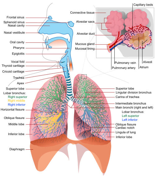
The Upper Respiratory Tract: The Air’s First Stop
The respiratory system can be divided into the upper and lower respiratory tracts. The upper respiratory tract includes the nose, nasal cavity, sinuses, pharynx (throat), and larynx (voice box). When we inhale, air first enters through the nose or mouth. The nose is the primary entry point, and it does more than just let air in. The nasal cavity is lined with mucous membranes and tiny hair-like structures called cilia. These work together to filter, warm, and humidify the incoming air, preparing it for its journey to the lungs.
The sinuses, hollow spaces in the bones of the face, also play a role. They produce mucus that helps moisten the air and trap dust and other particles. The pharynx is a passageway for both air and food. At its base is the larynx, which contains the vocal cords. When air passes over these cords, it creates the sounds of speech.
The Lower Respiratory Tract: Where Gas Exchange Happens
The lower respiratory tract consists of the trachea (windpipe), bronchi, bronchioles, and lungs. The trachea is a tube that carries air from the larynx to the bronchi. At its base, the trachea divides into two smaller tubes, the right and left bronchi, which lead to the right and left lungs, respectively.
Within the lungs, the bronchi branch into smaller and smaller tubes called bronchioles. At the end of each bronchiole are clusters of tiny air sacs called alveoli. This is where the magic of gas exchange takes place.
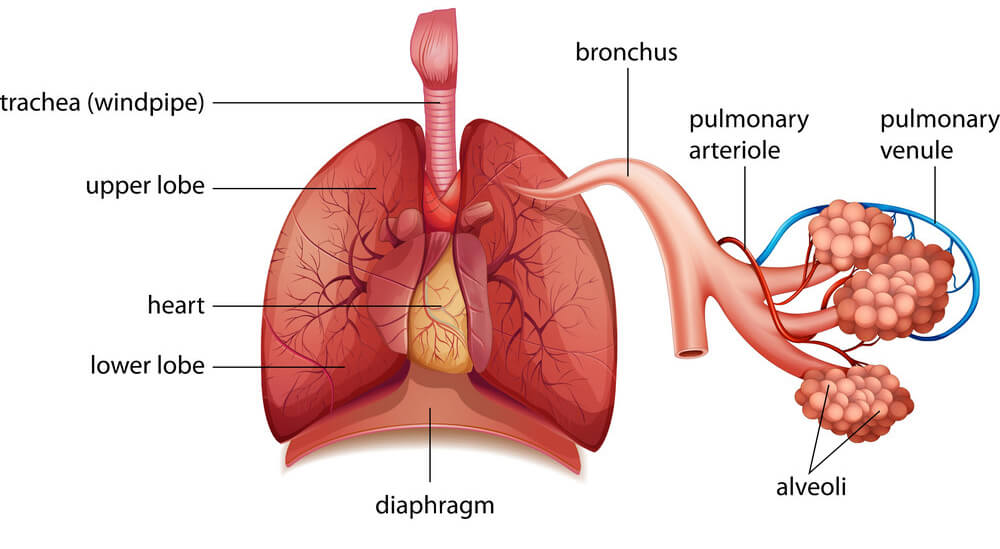
The alveoli are surrounded by a network of tiny blood vessels called capillaries. Oxygen from the air we breathe diffuses through the thin walls of the alveoli into the blood in the capillaries. At the same time, carbon dioxide from the blood diffuses into the alveoli to be exhaled. This process of gas exchange is crucial for delivering oxygen to all the cells in the body and removing the carbon dioxide they produce.
The Mechanics of Breathing: Inhale, Exhale
Breathing is a beautifully choreographed process that involves the diaphragm and other muscles. When we inhale, the diaphragm and intercostal muscles contract. This increases the volume of the chest cavity, creating a negative pressure that draws air into the lungs. When we exhale, these muscles relax, decreasing the volume of the chest cavity and forcing air out.
Breathing is usually an unconscious process controlled by the respiratory center in the brain stem. However, we can also control our breathing consciously, such as when we hold our breath or blow out candles on a birthday cake. The rate and depth of breathing can be influenced by factors such as exercise, emotions, and changes in blood pH levels.
The Respiratory System’s Defenses: Keeping the Airways Clear
With every breath, we inhale not just air but also dust, pollutants, and microbes. To protect the delicate tissues of the respiratory system, we have several lines of defense. The nose filters out large particles, while the mucus and cilia in the airways trap smaller particles and pathogens. The cilia constantly sweep the mucus and trapped debris upward to the throat, where it can be swallowed or coughed out.
The airways are also equipped with immune cells, such as macrophages, that engulf and destroy invading microbes. When harmful substances do make it past these defenses, inflammation, and increased mucus production occur as part of the immune response. While uncomfortable, coughing and sneezing help expel irritants and pathogens from the airways.
Respiratory Health and Homeostatic Imbalances
Maintaining the health of the respiratory system is vital for overall well-being. However, various factors can disrupt its normal functioning, leading to respiratory diseases. Smoking is a major risk factor, as it damages the cilia, increases mucus production, and causes chronic inflammation in the airways. This can lead to conditions such as chronic bronchitis and emphysema, collectively known as chronic obstructive pulmonary disease (COPD).
Asthma is another common respiratory condition characterized by inflammation and narrowing of the airways. Triggers such as allergens, cold air, and exercise can cause the airways to constrict, making breathing difficult. Proper management with medications and avoiding triggers are key.
Infections like the common cold, influenza, and pneumonia can also affect the respiratory system. While most infections are self-limiting, some can be severe, especially in vulnerable populations such as the elderly and those with compromised immune systems. Practicing good hygiene, getting vaccinated, and seeking prompt medical care when needed can help prevent and manage these infections.
The Breath-Body Connection: Posture, Stress, and Breathing
Did you know that your posture and emotional state can affect your breathing? Slouching compresses the chest cavity, reducing lung capacity. On the other hand, good posture allows for full chest expansion and deeper breaths. Stress and anxiety can also lead to rapid, shallow breathing, which can disrupt the balance of oxygen and carbon dioxide in the body.
Practicing deep, diaphragmatic breathing can help counteract these effects. By taking slow, deep breaths, we can activate the parasympathetic nervous system, inducing a relaxation response. This can lower heart rate, reduce blood pressure, and promote a sense of calm. Incorporating breathing exercises into your daily routine can help manage stress, improve posture, and support overall respiratory health.
Summary
From the moment we take our first breath to the last, the respiratory system works tirelessly to sustain life. Its intricate structure and precise mechanisms allow for the vital exchange of gases that every cell in our body depends on. By understanding how this system works and taking steps to protect it, we can breathe easier and support our overall health and well-being.